

 Advanced Technology


Cone Beam CT
Our practice utilizes state-of-the-art small-volume Cone Beam CT (CBCT) technology to provide highly detailed, three-dimensional images of your teeth and surrounding structures. Unlike traditional 2-D dental X-rays, CBCT allows us to view the tooth, bone, sinuses, and surrounding anatomy from multiple angles, giving us a more complete understanding of your individual anatomy.
This advanced imaging technology can be especially valuable when diagnosing complex endodontic conditions and planning treatment. By providing detailed 3-D images, CBCT can help us identify anatomy and conditions that may not be visible on traditional 2-D X-rays, allowing us to make more informed treatment decisions.
Our small-volume CBCT is designed to capture the area we need while using a low radiation dose, making it an important tool in providing precise, efficient, and patient-centered care.
gentlewave procedure
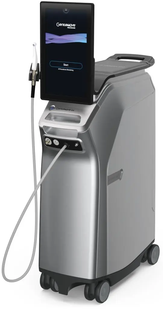
At our practice, we are proud to offer the GentleWave® Procedure, an innovative, minimally invasive technology for root canal treatment. Unlike traditional methods, GentleWave® uses advanced fluid dynamics and multisonic energy to thoroughly clean and disinfect even the most complex root canal systems—reaching areas conventional techniques often miss.
This state-of-the-art approach not only preserves more of the natural tooth structure but also promotes faster healing and improved outcomes, often in a single visit. For patients, that means a more comfortable experience with less post-operative discomfort.
moravision
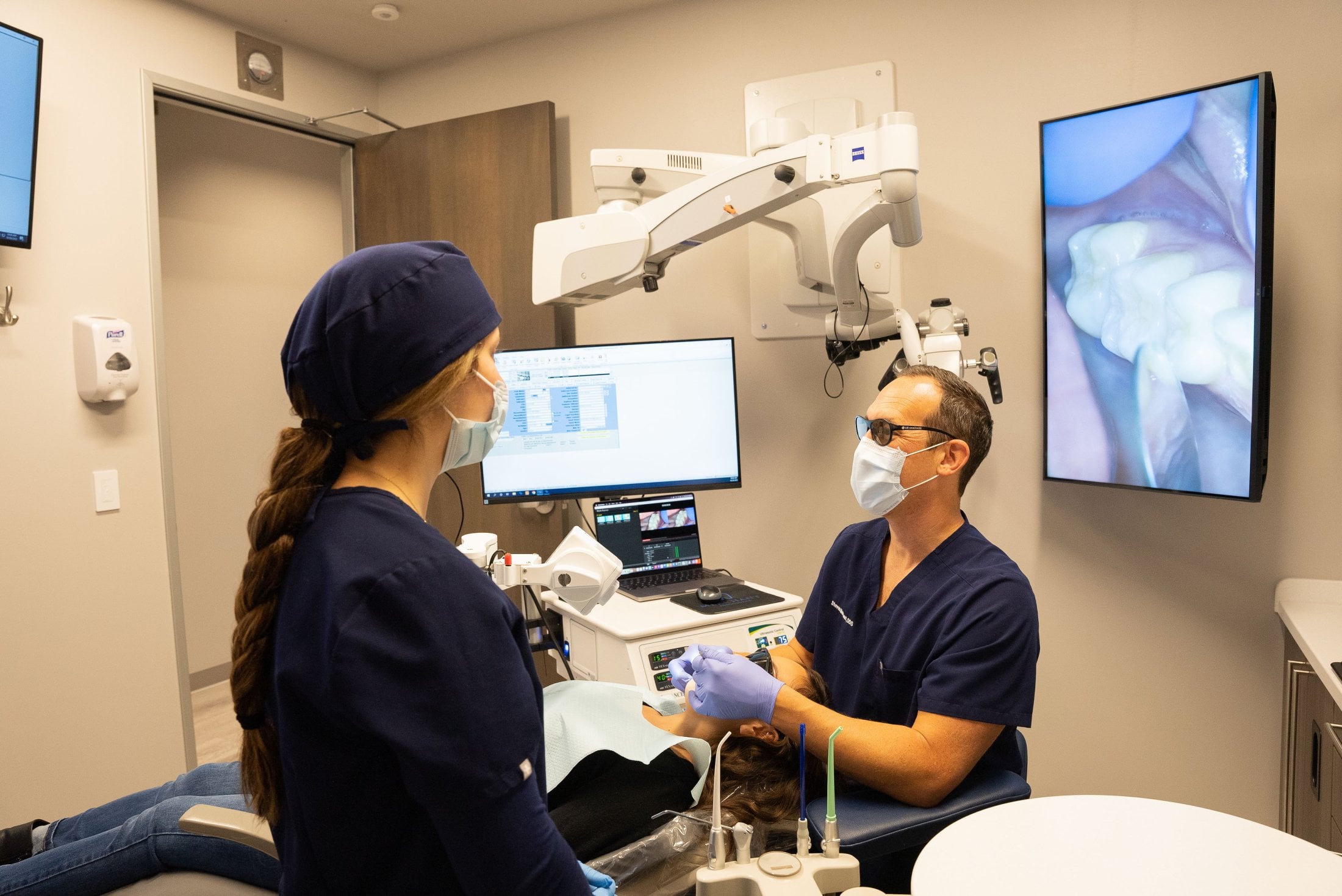
At Merchant Endodontics, we continually invest in advanced technology that enhances the precision, efficiency, and quality of your care. MoraVision® 3D is an advanced stereoscopic 3D dental microscopy system that provides real-time, high-definition visualization of the treatment area.
With up to 16× magnification, exceptional depth of field, and powerful coaxial LED illumination, MoraVision® 3D allows us to see fine anatomical details with remarkable clarity. This enhanced visualization can be especially valuable during complex endodontic procedures where precision is essential.
Unlike traditional microscopes that require the doctor to look through fixed eyepieces, MoraVision® 3D uses a high-resolution 3D camera and monitor. This allows for greater flexibility in positioning while maintaining a natural, ergonomic posture during treatment.
Benefits of MoraVision® 3D include:
- Real-time stereoscopic 3D visualization
- Up to 16× magnification
- Exceptional illumination and depth of field
- Flexible camera positioning
- Enhanced visualization of complex anatomy
- Improved ergonomics and operator comfort
- High-resolution digital documentation of procedures
By combining advanced 3D visualization with modern endodontic techniques, MoraVision® 3D helps us see more, work more precisely, and provide a higher level of care for our patients.
Surgical Microscopes

The surgical microscope has transformed the field of endodontic microsurgery, allowing endodontists to see details that may be difficult or impossible to visualize with the naked eye. At Merchant Endodontics, we have invested in high-quality Carl Zeiss surgical microscopes that provide exceptional magnification, illumination, and clarity during treatment.
In endodontics, precision matters. The ability to clearly visualize the smallest anatomical details allows us to perform treatment with greater accuracy and confidence. Our microscopes provide enhanced visualization of the treatment area, helping us identify structures and details that may otherwise go unseen.
We have also incorporated high-resolution digital photography into our microscopes, allowing us to document your tooth, diagnosis, and procedure with exceptional clarity and detail.
Digital Imaging

We carefully choose which and when radiographs are taken. There are many guidelines that we follow. Radiographs allow us to see everything we cannot see with our own eyes. Radiographs enable us to detect cavities, see signs of infection, determine bone level, and analyze the health of your bone. We can also examine the roots and nerves of teeth, diagnose lesions such as cysts or tumors, as well as assess damage when trauma occurs.
Dental radiographs are invaluable aids in diagnosing, treating, and maintaining dental health. Exposure time for dental radiographs is extremely minimal. We utilize Digital Imaging Technologies within the office. With digital imaging, exposure time is about 90 percent less when compared to traditional radiographs. Digital imaging can also help us retrieve valuable diagnostic information.
Digital imaging allows us to store patient images, and enables us to quickly and easily transfer them to your dentist, other specialists or insurance companies.